Learning center

Understanding mycoplasma testing

What are mycoplasmas?

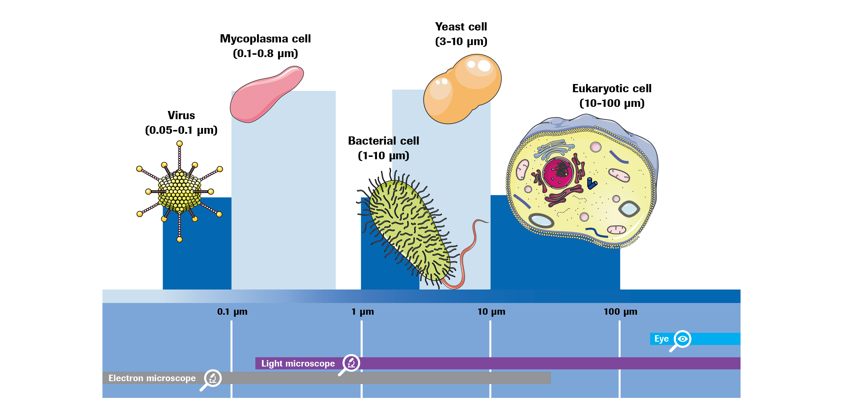
The term ‘mycoplasmas’ is often used as a trivial name for all members of the bacterial class Mollicutes (lat. mollis = „soft“, cutis = „skin“). Mollicutes are characterized by the lack of a cell wall and a small genome size (0.5 –2.2 megabase pair) with low GC (guanine-cytosine) content (20–40 mol%). Due to their small genome, mycoplasmas are host-dependent and live as commensals or infectious agents in or on a variety of hosts, including humans, other vertebrates, plants, and insects. These microorganisms can multiply under aerobic or anaerobic conditions. They have a pleomorphic cell morphology, with the exception of spiroplasmas, which have a spiral shape, and some mycoplasmas of the genus Mycoplasma, which have a flask-like shape due to a terminal (tip) structure (Mycoplasma gallisepticum, Mycoplasma pneumoniae).

Depending on species, mycoplasmas can grow in liquid media, either as single cells (Mycoplasma arthritidis) or in aggregates (Acholeplasma laidlawii, Mycoplasma pneumoniae, Mycoplasma fermentans). The lack of a cell wall makes mycoplasmas resistant to cell wall-targeting antibiotics such as penicillin. Furthermore, some mycoplasmas can form biofilms on solid surfaces in liquid media, such as glass or plastic surfaces, which provides another level of resistance, namely to disinfecting agents and environmental stress conditions.

Mycoplasma in cell cultures

Mycoplasma (contamination) in cell cultures

In addition to their growing clinical significance, mycoplasmas have gained great attention in the context of cell cultures. As they naturally reside in plant and animal tissues, every cell culture medium containing plant- or animal-derived supplements is prone to contamination by mycoplasmas. Due to their small size of only 0.1–0.8 μm on average, and their variable shape resulting from the missing cell wall, mycoplasmas can pass through standard sterilizing filters and enter cell cultures with culture media or raw material-derived additives.


The two most common sources of contamination

The two most common sources of contamination are laboratory personnel and already contaminated cell cultures, from which the contaminant is passed on by cross-contamination. Since mycoplasmas are not visible with standard light microscopy setups and usually barely affect the obvious state of the cell culture, they often remain undetected. Nevertheless, they impact cell growth and metabolism, and consequently, the therapeutic proteins expressed by host cells.

Mycoplasma species frequently, potentially or occasionally detected in cell cultures and in biopharmaceutical processes

View Full Table

Mycoplasma species frequently, potentially or occasionally detected in cell cultures and in biopharmaceutical processes

Mycoplasma species which are frequently, occasionally or potentially detected in cell cultures and in biopharmaceutical processes.

Mycoplasma species which are frequently, occasionally or potentially detected in cell cultures and in biopharmaceutical processes.

Mycoplasma species Primary isolation source (relevant for products where raw materials of the following origins are used) Frequent cell culture contaminant based on published reports Potential contamination source
Acholeplasma laidlawii Bovine, prcine, avian, plant Yes Other cell line, bovine sera, nutrient broth powders
Mycoplasma arginini Bovine, ovine, caprine, porcine Yes Other cell line, bovine sera
Mycoplasma bovis Bovine Yes Other cell line, bovine sera
Mycoplasma fermentans Human Yes Other cell line, personnel
Mycoplasma gallisepticum Avian No Other cell line, embryonated eggs
Mycoplasma hyorhinis Porcine Yes Other cell line, porcine trypsin
Mycoplasma orale Human Yes Other cell line, personnel
Mycoplasma salivarium Human Yes Other cell line, personnel
Mycoplasma synoviae Avian No Other cell line, embryonated eggs
Spiroplasma citri Plant No Other cell line

Mycoplasmas in Manufacturing Processes of Biopharmaceuticals and ATMP

Especially in the biopharmaceutical and cell therapy industry, the effects of mycoplasma contamination are devastating, as entire production batches must be discarded and the manufacturing plant must stop production. International regulatory authorities have published guidelines to demonstrate that biological products intended for preventive or therapeutic clinical use and prepared in cell culture substrates must be free of mycoplasmas to ensure product safety, purity and potency. Therefore, early detection of mycoplasmas is essential for smooth processes in manufacturing of biopharmaceutical and cell therapy products. 

Advanced therapy medicinal products (ATMP) are a new class of therapeutics that are based on genes (gene therapy), somatic cells (cell therapy), or tissue (tissue engineering). These advanced therapies herald novel treatments of a number of diseases and thus, a huge potential for patients is expected. Autologous cell therapy, for instance, typically involves cell dissociation from an individual patient, cell culture outside the human body, and subsequent injection of cells back into the patient.

Biopharmaceuticals (also known as biologics) are the ‘classical’ medicinal products manufactured in and extracted from biological sources such as bacteria, yeast, mammalian cell lines, or mammals. Vaccines, purified blood components and recombinant proteins fall into this category. They can consist of nucleic acids proteins, sugars and complex combinations of these and are either identical or similar to molecules naturally occurring in the human body.

Mycoplasma detection


Testing points for mycoplasma contamination in the manufacturing process of biopharmaceuticals

After raw materials have been tested for contamination (grey loop), and solutions like buffer and media have been applied to working cultures, it is necessary to check for contamination (blue loop), as mycoplasma contamination can also be introduced by the cell line and lab staff. It is also recommended to carry out in-process controls during the seed culture and the actual fermentation (light purple loops). The final and prescribed test point is the endpoint of the fermentation, the harvest (purple loop). Once the mycoplasma-free state of the harvest has been proven, further test points are usually no longer necessary since the purification of the products is carried out without living organisms. 

Mycoplasma detection

The picture depicts common testing points in the manufacturing process of ‘classical’ biopharmaceuticals. Given these multiple checkpoints, numerous different methods for mycoplasma testing have been developed and will be covered in the following section.

Methods overview

Non-compendial testing methods


Non-compendial tests for mycoplasma detection often lack the sensitivity to detect the level of contamination in a sample that is required by regulatory monographs.
Moreover, results are sometimes difficult to interpret if a contamination is at low level. These tests are prone to giving false-negative results.

Direct DNA staining

Direct staining of cultures with a DNA (Deoxyribonucleic acid)-specific fluorescent dye is sensitive, but not recommended for the purpose of detecting mycoplasma contaminations. Although the test reliably detects heavily contaminated cultures, interpretation of low-grade contaminations is often difficult because DNA from the cell culture may give rise to small points of fluorescence that can mimic mycoplasmas.

Enzyme-based method

Enzyme-based assays are selective biochemical tests that exploit the activity of mycoplasma enzymes. A prerequisite for such a test to be applicable for routine mycoplasma testing is that the enzymatic activity measured must ideally be ubiquitous among mycoplasmas, but missing in the eukaryotic cell matrix.

Mycoplasma PCR-ELISA

An application that combines PCR with a subsequent ELISA (enzyme-linked immunosorbent assay) is the mycoplasma PCR ELISA, a photometric enzyme immunoassay that detects PCR amplified mycoplasma DNA in cell culture. During the PCR reaction, digoxigenin-labeled nucleotides are incorporated into the amplicons, allowing their detection in a subsequent ELISA assay. The mycoplasma PCR-ELISA test is claimed to have a detection sensitivity of 1–3 mycoplasma “particles” for particular mycoplasma species (e.g. ,M. fermentans and A. laidlawii).22 However, since for others the Limit of Detection (LOD) was 1000 “particles” per ml sample, the test does not fulfill the requirements of the EP regulatory guideline as compendial test for mycoplasma detection.

Compendial testing methods


These methods are, on one hand, based on conventional microbiological culture procedures using liquid media and agar media and, on the other hand, based on rapid molecular techniques.

Culture method
 

Traditional culture methods were used well before today’s molecular techniques and are still found in regulatory and compendial protocols throughout the world (formulated in the EP, USP and JP regulatory guidelines). The Culture Method is based on the targeted cultivation of mycoplasmas in culture media that promote mycoplasma growth. A sample to be tested is inoculated into the liquid mycoplasma culture media and onto agar media, and mycoplasma growth is promoted by microaerophilic incubation conditions, such as 36 ± 1°C, 5.5 ± 0.5% CO2, 3 ± 1% O2 and 90 ± 5% relative humidity. Subcultivation from the liquid cultures onto agar plates is carried out up to 21 days after the initial inoculation. On the agar medium, mycoplasmas develop microscopic colonies (< 100–400 μm diameter).

NAT-based method
 

NAT-based methods include all tests based on nucleic acid detection, often performed by PCR.

Indicator cell culture method
 

The Indicator cell culture method is normally carried out with Vero or 3T3 cell lines, although the use of a production cell line that is equivalent in effectiveness for detecting mycoplasmas is also accepted by the EP regulatory guideline. The indicator cell culture is inoculated with the sample and incubated at 35–38°C until grown to confluence. For positive controls, the indicator cell line is also inoculated with the type strain CH19299T of M. orale and the M. hyorhinis cultivar alpha reference strain DBS 1050 with and without the presence of the test sample. Before staining, the subculture is fixed with a suitable fixing solution and stained with a fluorescent dye that binds to DNA. The presence of mycoplasmas is characterized by a spherical fluorescence pattern on the cell surface and by strong fluorescence in the surrounding areas. Mitochondria in the cytoplasm are also stained, but are easily distinguished from mycoplasmas. The test is invalid if the positive controls do not show fluorescence typical for mycoplasmas or if the negative control shows fluorescence typical for mycoplasmas.

Learn about our mycoplasma testing solutions
View products

Regulatory disclaimers are listed on the respective product pages.